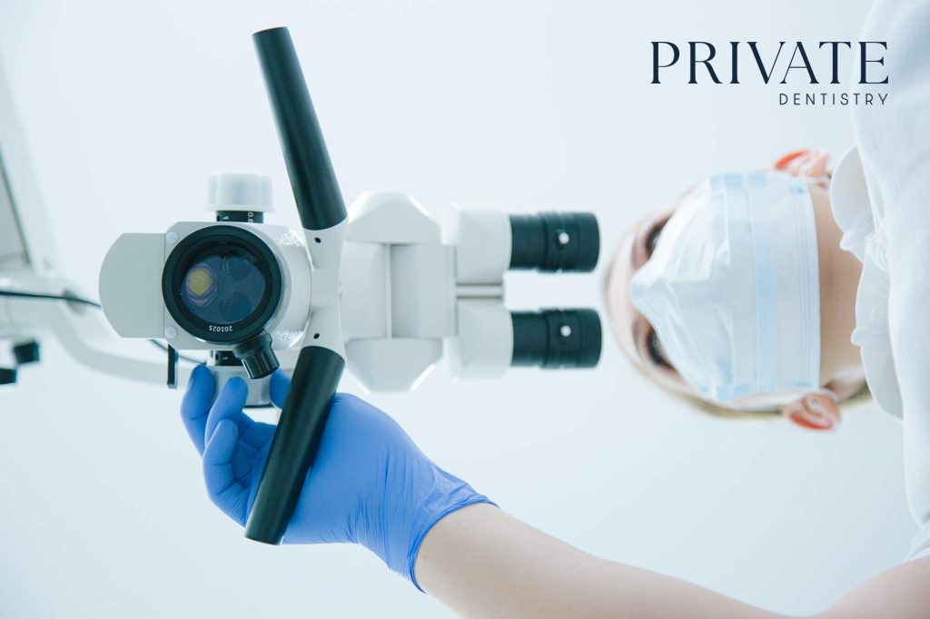
Brooke Shipp explains how microscopy has revolutionised her clinical practice by allowing patients to visualise gum disease.
As dental professionals, we often speak to patients about plaque, biofilm and periodontal disease, but how often do they really understand what that means? Despite our best efforts with diagrams, charts and models, many patients still struggle to grasp the invisible reality of gum disease. That’s where high-magnification microscopy has revolutionised my clinical practice.
Making the invisible visible
Recently, I integrated a microscope into my periodontal assessment workflow. This tool allows me to collect a plaque sample from the tooth surface and display a live image of the bacteria directly onto a screen for the patient to view. This real-time visualisation immediately shifts the dynamic of the consultation. The patient is no longer hearing abstract explanations they’re witnessing the pathogens moving on screen, in their own mouth.
One patient, in particular, had been struggling with bleeding gums and chronic halitosis, yet was inconsistent with home care and unsure why treatment was necessary. Once I placed their plaque sample under the microscope and projected the live video, their reaction was immediate: shock, curiosity, and finally, motivation.
I used the opportunity to explain how the gram-negative anaerobic bacteria I observed are linked to periodontal destruction, inflammation, and tissue breakdown. This visual connection between what they saw and what they felt made the disease real and urgent to them.